Diese Doppelhelix — antiparallele Stränge, farbcodierte Basenpaare, Furchen-Hinweise, ein Replikations-Einschub — stammt aus einem einzigen Prompt mit rund 40 Wörtern. Im Folgenden finden Sie 25 Biologie-Prompts auf demselben Niveau, nach Themen geordnet, jeder mit seiner Beschriftungsliste und dem konkreten Punkt, den es zu prüfen gilt. Fügen Sie sie direkt in den Generator ein oder passen Sie sie an Ihr genaues Thema an.
Biologie-Voreinstellungen öffnen · Vollständige Arbeitsablauf-Anleitung · Galerie mit 30 Beispielen
Hinweis zur Sprache: Alle Prompts bleiben unverändert auf Englisch, weil die auf den Abbildungen gerenderten Beschriftungen englisch erscheinen und kurze Großbuchstaben-Labels am zuverlässigsten gerendert werden.
So lesen Sie dieses Paket: Jeder Prompt folgt einer Grammatik — zuerst das Thema, dann die Panels, dann die Beschriftungen in Anführungszeichen, dann ein Titel. Die zitierten Beschriftungen sorgen dafür, dass Text korrekt gerendert wird; die benannten Panels sorgen dafür, dass das Layout eine Abbildung wird und keine Wanddekoration. Passt ein Thema unten nicht exakt zu Ihrer Unterrichtsstunde, ändern Sie die Themenzeile und behalten die Struktur.
Zellen
1. DNA-Doppelhelix
An elegant structural figure of the DNA double helix: two antiparallel strands,
base pairs as rungs with the four bases color-coded, a major and minor groove
callout, and one unzipping replication inset. Label "STRAND", "BASE PAIR", "MAJOR
GROOVE", "MINOR GROOVE". Title: "THE DOUBLE HELIX".
Vor Verwendung prüfen: Die Stränge müssen antiparallel verlaufen (5'→3' in der einen, 3'→5' in der anderen Richtung); A-T-Paare haben zwei Wasserstoffbrücken-Striche, G-C drei — prüfen Sie, ob der Einschub sie nicht vereinheitlicht hat.
2. Replikationsgabel der DNA
A DNA replication fork with the parental double helix opening into a leading strand
and a lagging strand with Okazaki fragments, helicase at the fork and DNA polymerase
on the new strand, synthesis in the 5' to 3' direction. Label "HELICASE", "LEADING
STRAND", "LAGGING STRAND", "OKAZAKI FRAGMENT", "DNA POLYMERASE".
Title: "THE REPLICATION FORK".

Vor Verwendung prüfen: Die Okazaki-Fragmente des Folgestrangs müssen von der Gabel weg zeigen; beide neuen Stränge werden 5'→3' synthetisiert — zeigt die Zeichnung etwas anderes, generieren Sie mit dieser Randbedingung erneut.
3. Zellorganellen der Tierzelle
A large cutaway animal cell with its organelles arranged realistically, each
organelle shown once with a short functional caption beside it in a small grid
around the cell. Label "NUCLEUS", "MITOCHONDRION", "ROUGH ER", "GOLGI",
"LYSOSOME". Title: "THE ANIMAL CELL".

Vor Verwendung prüfen: Das raue ER liegt dicht am Zellkern; das Zentrosom liegt nahe dem Kern, nicht an der Membran. Zwei Zellen im Vergleich:
4. Tierzelle gegen Pflanzenzelle
A paired cutaway comparison of an animal cell and a plant cell at matching scale,
with shared organelles in the middle band and unique structures at the outer edges,
plus a small legend for the color coding. Label "NUCLEUS", "MITOCHONDRION", "CELL
WALL", "CHLOROPLAST", "VACUOLE". Title: "ANIMAL VS PLANT".

Vor Verwendung prüfen: Die große Zentralvakuole und die Zellwand gehören allein auf die Pflanzenseite; prüfen Sie, ob die Farblegende zum tatsächlich Gezeichneten passt.
5. Vier spezialisierte Tierzellen
A comparative row of four specialized cells at matching scale: a neuron with
dendrites and long axon, a red blood cell as a biconcave disc, a sperm cell, and a
macrophage spreading pseudopods, each with two functional labels.
Title: "CELL SPECIALISTS".

Vor Verwendung prüfen: Größenangaben sind hier heikel — kennzeichnen Sie, dass die Reihe nicht maßstabsgetreu ist (ein Erythrozyt misst rund 7 µm, ein Axon kann einen Meter erreichen), solange Sie keinen Maßstabsbalken ergänzen.
Photosynthese und Pflanzenbiologie
6. Lichtreaktion der Photosynthese (Z-Schema)
A Z-scheme figure of the light reactions: photosystem two splitting water and
sending electrons through plastoquinone, cytochrome, plastocyanin to photosystem
one, ferredoxin reducing NADP, and the proton gradient feeding ATP synthase.
Label "PSII", "PSI", "PLASTOQUINONE", "NADP", "ATP SYNTHASE".
Title: "Z SCHEME".

Vor Verwendung prüfen: Richtung des Elektronenflusses PSII → PSI; Wasser wird am PSII gespalten (dort wird Sauerstoff frei), die ATP-Synthase sitzt in der Thylakoidmembran mit Protonen darüber im Lumen.
7. Protonenverkehr im Chloroplasten
A chloroplast figure centered on the thylakoid membrane: protons pumped into the
lumen by the electron transport chain flowing back out through ATP synthase,
stroma-side NADP reduction, and one granum stack inset showing where all this sits.
Label "LUMEN", "STROMA", "PROTON", "ATP SYNTHASE", "NADP".
Title: "PROTON TRAFFIC".
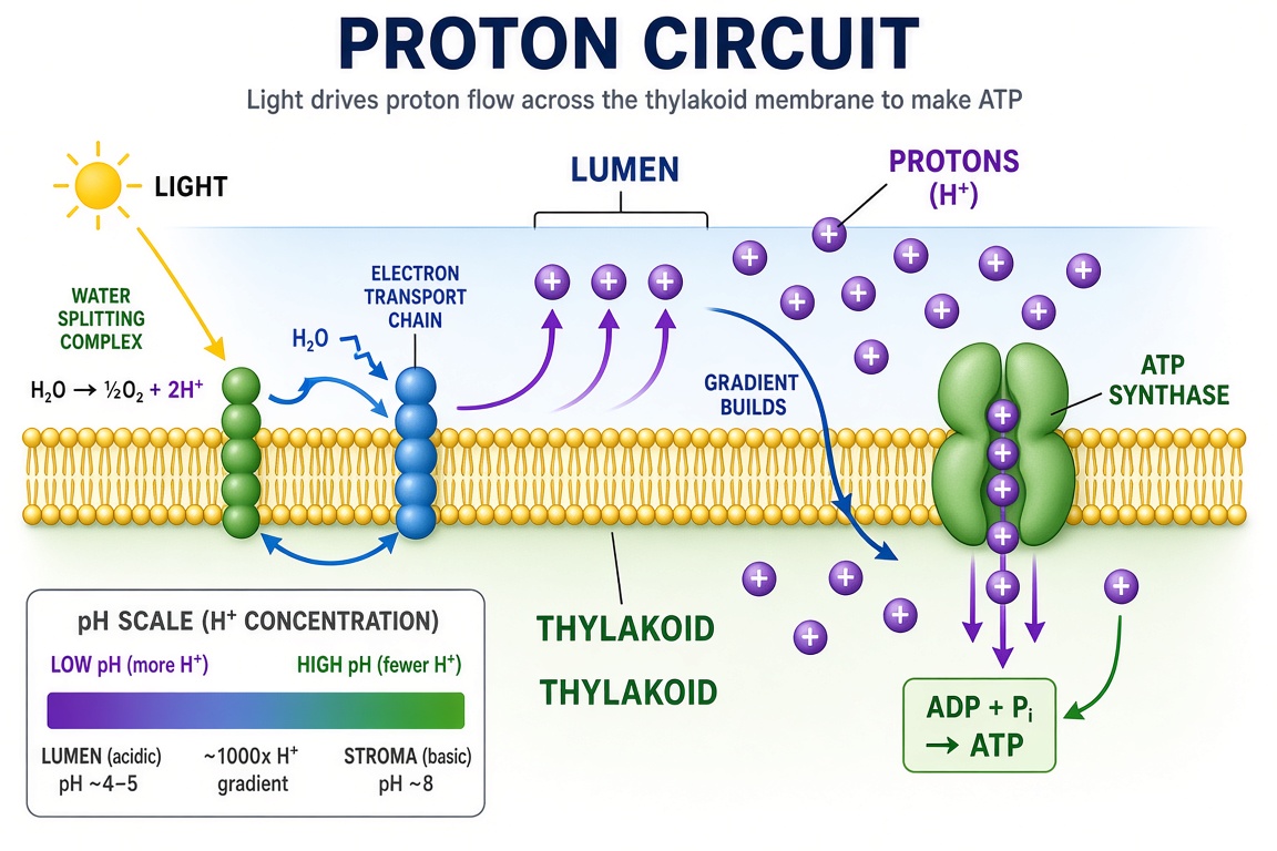
Vor Verwendung prüfen: Die Seiten von Lumen und Stroma — Protonen reichern sich im Lumen an; der Calvin-Zyklus gehört ins Stroma, nicht ins Lumen.
8. Trockenheitsantwort der Pflanze
A plant-physiology figure of drought response: roots sensing dry soil, abscisic
acid traveling up the xylem, guard cells closing stomata, leaf rolling and
osmoprotectant accumulation, with a healthy versus stressed plant pair.
Label "ABA", "GUARD CELL", "STOMATA", "OSMOPROTECTANT". Title: "SURVIVING DRYNESS".
Vor Verwendung prüfen: Beim gestressten Exemplar sind die Stomata geschlossen (Schließzellen schlaff), nicht offen; der ABA-Pfeil verläuft von der Wurzel über das Xylem zum Blatt.
Genetik und Molekularbiologie
9. CRISPR-Cas9-Mechanismus
A gene-editing mechanism figure: the Cas9 protein as a bilobed clamp, guide RNA
threaded inside base-pairing with the target DNA, the HNH and RuvC domains cutting
both strands, and a repaired insert outcome panel. Label "CAS9", "GUIDE RNA",
"TARGET DNA", "CUT SITE", "INSERT". Title: "GENE EDITING".
Vor Verwendung prüfen: Die Guide-RNA paart mit dem Zielstrang; die PAM-Sequenz liegt auf dem Nicht-Zielstrang unmittelbar hinter der Schnittestelle.
10. Antikörper-Antigen-Bindung
A molecular figure of an antibody with its two heavy and two light chains, the
antigen-binding sites gripping a small antigen, and one magnified inset of the
paratope-epitope interface. Label "HEAVY CHAIN", "LIGHT CHAIN", "PARATOPE",
"EPITOPE". Title: "ANTIBODY STRUCTURE".

11. Bakteriophage T4
A structural figure of the T4 bacteriophage with its icosahedral head, contractile
sheath, tail fibers and base plate, plus one inset showing the tail injecting DNA
into a bacterial envelope. Label "HEAD", "SHEATH", "TAIL FIBER", "BASE PLATE",
"DNA". Title: "T4 PHAGE".

Vor Verwendung prüfen: Im Infektions-Einschub docken die Schwanzfasern vor der Kontraktion der Scheide an.
12. Aggregationsstufen von Amyloid-beta
A four-stage molecular figure of peptide aggregation: free monomers, small oligomer
rings, a growing fibril strand shown as a twisted beta-sheet ladder, and a dense
senile plaque mass with microglia reacting at the edge. Label "MONOMER", "OLIGOMER",
"FIBRIL", "PLAQUE". Title: "AGGREGATION".
Vor Verwendung prüfen: Der Fibril-Querschnitt ist ein Beta-Faltblatt-Stapel; wirken die Sprossen wie eine Alpha-Helix, generieren Sie erneut.
Mikrobiologie und Labor
13. 96-Well-Platten-Layout
A top-down layout of a 96-well microplate set up for a dose-response assay: eight
rows by twelve columns, control wells in the outer columns, a serial dilution
gradient across the middle, and triplicate rows. Label "CONTROL", "DILUTION
SERIES", "REPLICATES" and add a small color-concentration legend.
Title: "PLATE LAYOUT".
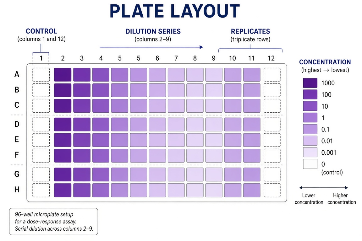
Vor Verwendung prüfen: Der Verdünnungsgradient muss über die Spalten in einer Richtung verlaufen; Dreifachansätze sind Zeilen, nicht Spalten.
14. RT-qPCR-Arbeitsablauf
A bench workflow figure for a quantitative PCR run: template and master mix on ice,
plate setup with standards and controls in a layout grid, thermal cycling profile
with annealing step, and melt-curve verification at the end. Label "MASTER MIX",
"STANDARDS", "THERMAL CYCLE", "MELT CURVE". Title: "THE QPCR RUN".

15. Sandwich-ELISA
A four-step laboratory workflow for a sandwich ELISA from coating the plate through
blocking and sample incubation to detection, each step as a small labeled panel
connected by arrows. Label "COATING", "BLOCKING", "INCUBATION", "DETECTION".
Title: "SANDWICH ELISA WORKFLOW".

Vor Verwendung prüfen: Zuerst der Capture-Antikörper unten, der Detektions-Antikörper oben — die Reihenfolge des Sandwiches ist der häufigste Fehler im Unterricht.
Entwicklung und Systeme
16. Bioprinting eines Gewebegerüsts
A figure of extrusion bioprinting: a printhead depositing parallel bioink strands
onto a grid scaffold, cells visible inside the strands in a magnified inset, and
the finished layered tissue construct on the right. Label "PRINTHEAD", "BIOINK",
"CELLS", "SCAFFOLD", "CONSTRUCT". Title: "BIOPRINTING".
17. Stammbaum der Blutzellen
A classic hematopoiesis tree: the hematopoietic stem cell at the root branching
through progenitors into red cells, platelets, granulocytes, monocytes and
lymphocytes, each mature cell drawn at the leaf. Label "STEM CELL", "PROGENITOR",
"RED CELL", "PLATELET", "LYMPHOCYTE". Title: "BLOOD LINEAGES".
Acht schnelle Varianten
Thema tauschen, Grammatik behalten:
| Nr. | Thema | Einzeilige Änderung |
|---|---|---|
| 18 | Prokaryot gegen Eukaryot | "A paired comparison of a bacterial cell and a eukaryotic cell… Label 'NUCLEOID', 'RIBOSOMES', 'PILI', 'NUCLEUS'" |
| 19 | Mitose-Überblick | "A five-panel row of mitosis stages: prophase, prometaphase, metaphase, anaphase, telophase… Label each panel" |
| 20 | Crossing-over der Meiose | "A tetrad with chiasmata crossing over between homologous chromosomes… Label 'TETRAD', 'CHIASMA', 'RECOMBINANT'" |
| 21 | Enzymkinetik | "A lock-and-key figure plus a Michaelis-Menten curve inset… Label 'SUBSTRATE', 'ACTIVE SITE', 'VMAX', 'KM'" |
| 22 | Stickstoffkreislauf | "A four-reservoir cycle figure: atmosphere, soil, plants, decomposers… Label 'N2', 'NITRIFICATION', 'ASSIMILATION', 'DENITRIFICATION'" |
| 23 | Neuronale Synapse | "A chemical synapse with vesicles fusing, neurotransmitters crossing the cleft, receptors on the postsynaptic side… Label 'VESICLE', 'CLEFT', 'RECEPTOR'" |
| 24 | Zellmembran | "A fluid-mosaic membrane cross-section with phospholipids, channel protein, glycoprotein… Label 'PHOSPHOLIPID', 'CHANNEL', 'GLYCOPROTEIN'" |
| 25 | Transkription | "A DNA strand opening with RNA polymerase transcribing mRNA, one gene with promoter and terminator… Label 'PROMOTER', 'RNA POLYMERASE', 'mRNA'" |
Prüfliste für wissenschaftliche Abbildungen
- [ ] Sind alle Beschriftungen korrekt geschrieben?
- [ ] Sind alle angeforderten Strukturen vorhanden?
- [ ] Sind Positionen und Proportionen gegenüber der Referenz plausibel?
- [ ] Zeigen die Pfeile in die richtige Richtung?
- [ ] Sind die Phasen in der richtigen Reihenfolge dargestellt?
- [ ] Wurde das Ergebnis mit einer verlässlichen Quelle abgeglichen?
Nächste Schritte
Lassen Sie einen der Prompts oben in den Biologie-Voreinstellungen laufen und gleichen Sie das Ergebnis vor der Unterrichtsstunde mit dem Schulbuch ab. Vertiefende Lektüre: Anatomie-Prompts für Herz, Hirn und Neuronen, botanische Prompts und zoologische Prompts. Was im Allgemeinen schiefgeht und wie man es per Prompt korrigiert, beschreibt 12 typische KI-Fehler bei wissenschaftlichen Illustrationen.