What Is Medical Illustration?
Medical illustration is the visual side of clinical and biological communication. It draws the body — organs, tissues, systems, surgical steps — so that students, patients, and clinicians can see structure and process clearly. A heart cutaway that shows the chambers and valves in their correct spatial relationship does more teaching in one image than a paragraph can, and that is the job medical illustration exists to do.
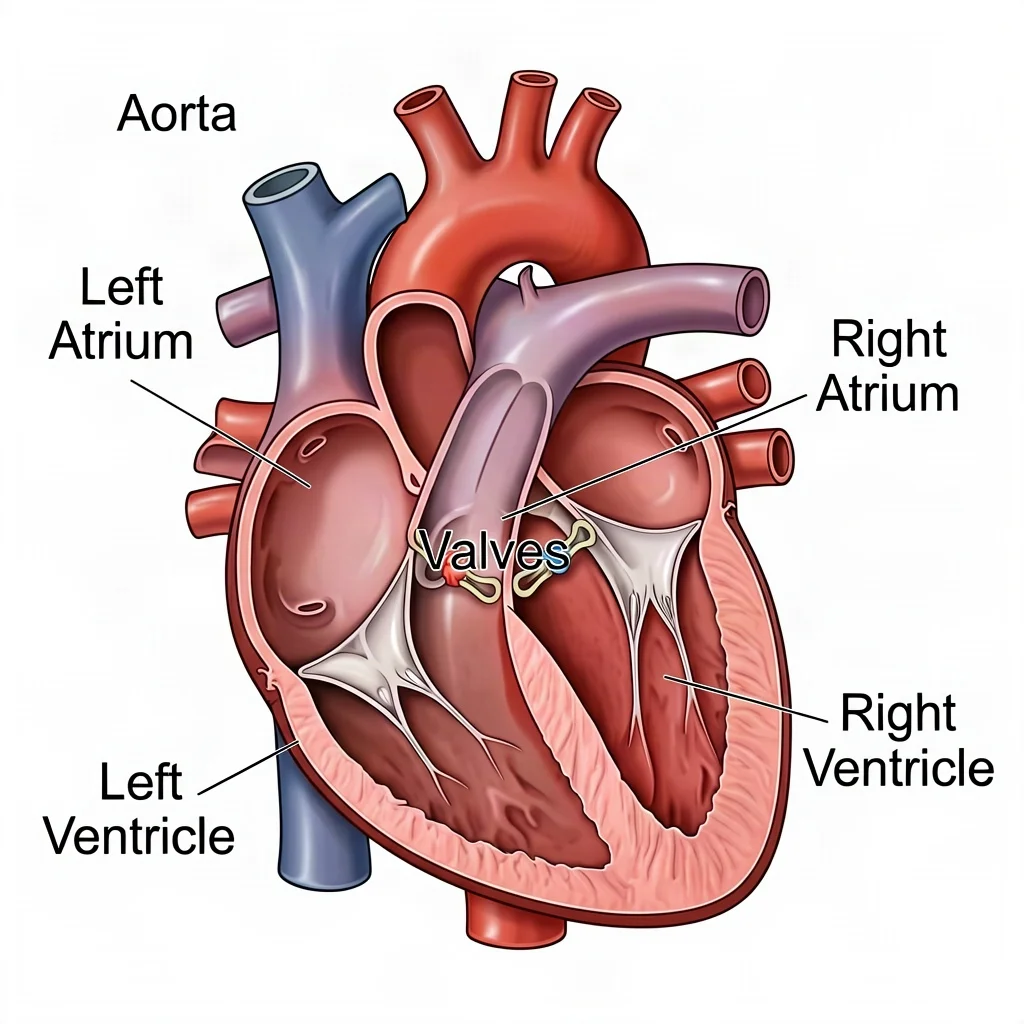
You will find medical illustration in textbooks, journal articles, patient-education handouts, trial exhibits, and surgical training. Where a photograph is too literal or too graphic, and a diagram is too abstract, medical illustration lands in between — clear, accurate, and labeled.
What Is Medical Illustration Used For?
The work splits into a few recurring jobs.
- Anatomy figures show structure — where things are and how they relate, from a whole-body schematic to a single receptor.
- Process figures show what happens — a disease progressing, a procedure unfolding, a drug moving through the body.
- Instructional figures show what to do — exercises, device use, post-operative care.
- Molecular and research graphics explain mechanisms at scales no camera can reach: cell signaling, DNA replication, drug binding.
In every case the image has to be readable, correctly proportioned, and labeled in plain clinical language. A medical illustration that looks right but is topologically wrong is worse than useless in a clinical setting, because it teaches a falsehood with confidence.
The Conventions That Make a Clinical Figure Readable
Good medical illustration follows habits the eye relies on.
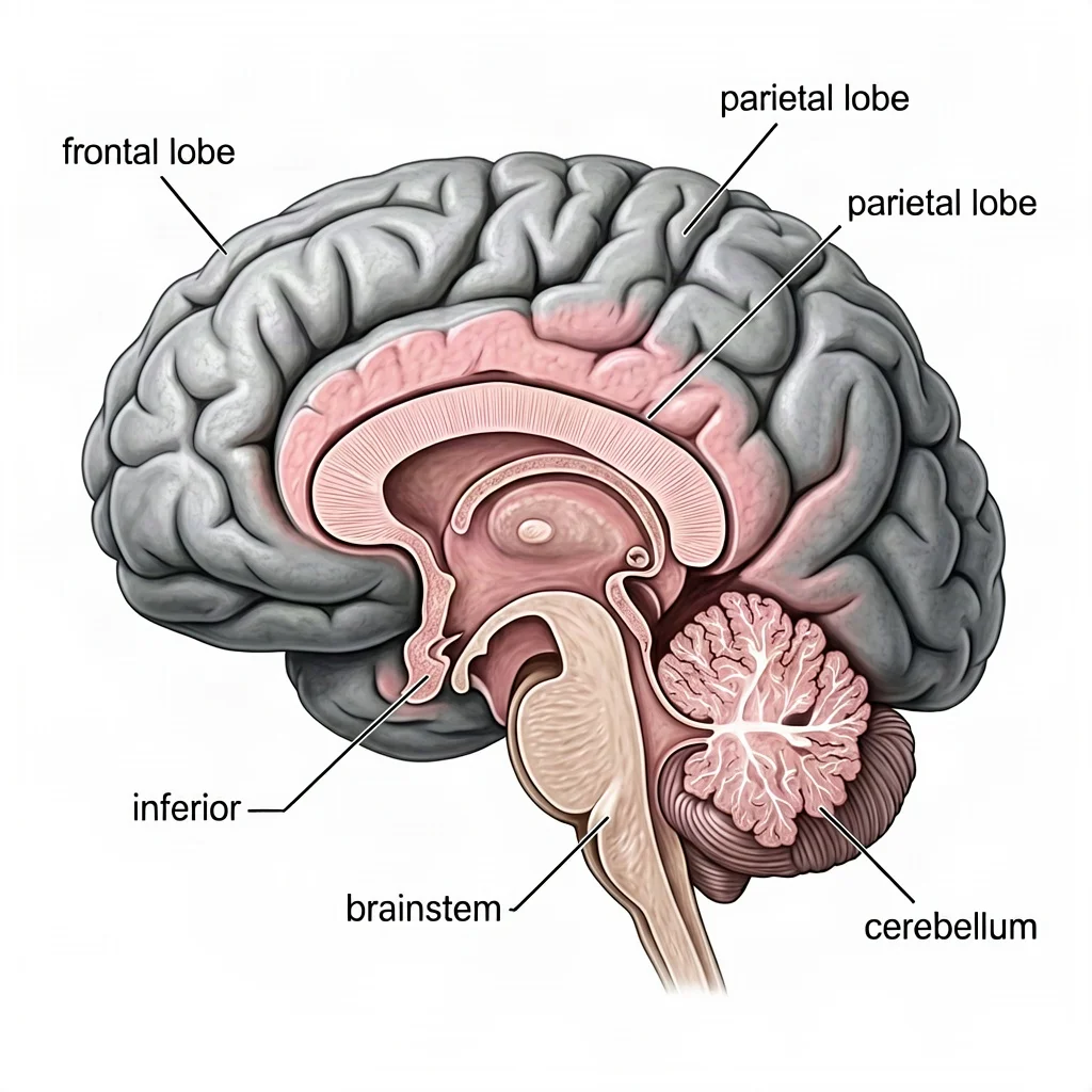
- Color coding with restraint. Arteries red, veins blue, nerves yellow, bone off-white — consistent and limited, so the reader maps color to meaning without thinking. The coding only works if it is never broken mid-figure.
- Standard views and orientations. Anterior, lateral, sagittal, cross-section — named views mean every reader interprets the figure from the same angle. Directional terms (superior, inferior, anterior, posterior) anchor the description.
- Correct spatial relationships. Chambers, vessels, and layers sit where they actually do. A figure earns trust by being right topologically, not just visually pleasing.
- Cutaways and layering. The body is opaque, so medical illustration routinely cuts it open, removes overlying structures, or renders them ghosted to expose what matters — an arrangement no dissection photo can match.
- Labels with leader lines. Each named part gets a thin line to its structure, arranged so labels do not cross or crowd.
How to Approach an Anatomical Figure
- Pick the view that best shows the target structure — anterior, sagittal, cross-section.
- Block in proportion and orientation from a trusted anatomical reference.
- Render the target structures accurately — layers, connections, branching.
- Add clinical color coding, kept minimal and consistent.
- Label the structures a reader needs to identify.
- Verify against a reference before you rely on it.
How Accurate Does Medical Illustration Need to Be?
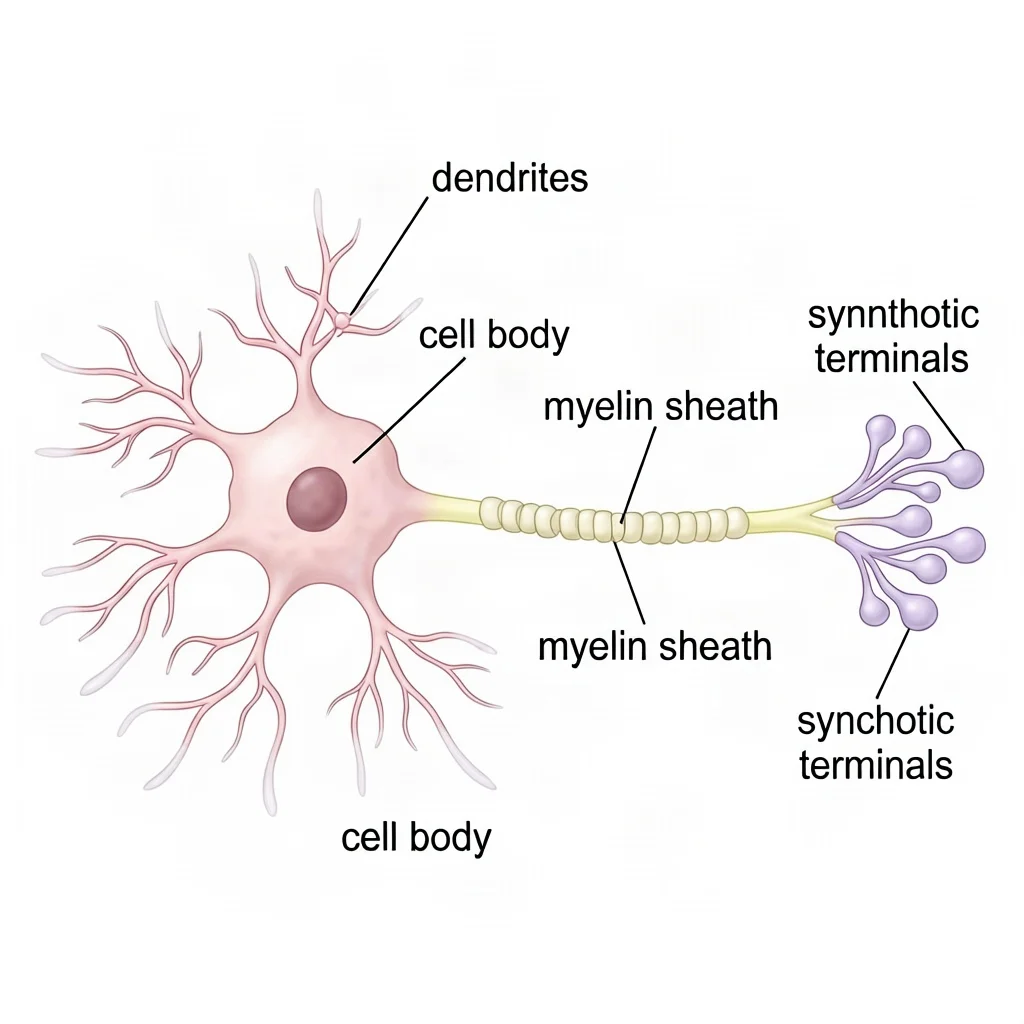
It depends on the audience, and the bar is highest where the stakes are. A medical illustration for a peer-reviewed paper or a surgical plan is checked structure by structure against established anatomy. A medical illustration for a patient handout or a lesson can simplify, as long as it never misleads — a patient following a post-operative figure has to recognize their own body in it. The discipline's reputation rests on that line: simplify, but do not falsify.
Where Medical Illustrators Work
The profession has a documented founding: Max Brödel, hired at Johns Hopkins at the end of the 19th century, drew for its surgeons and, in 1911, directed the first academic department of medical illustration. His students carried the model to other institutions, and the field's center of gravity has stayed in hospitals and universities ever since.
Today the profession is small and spread across a handful of employer types. Many medical illustrators work at hospitals and academic medical centers, producing figures for research publications, surgical training, and patient materials. Others work in pharmaceutical and biotech companies, building mechanism-of-action visuals for drugs and devices. Medical-legal firms hire illustrators to make trial exhibits of injuries and procedures that a jury can follow. Publishers, animation and simulation studios, and agencies serving all of the above employ more. A large share of the field is freelance or runs a small studio, serving several of these clients at once — which is why the skill set usually includes business practice alongside anatomy and rendering.
The day-to-day work varies as much as the employers. One week may be a series of patient-education panels that must read at a sixth-grade level; the next, a surgical sequence checked against operative video for a training module; the next, a journal figure that compresses a study's central mechanism into one panel. What every job shares is the review loop: content arrives from clinicians and researchers, the illustrator translates it, and the experts verify the result before it ships.
Training, Certification, and What the Work Pays
The standard entry route in North America is a graduate program accredited by the Association of Medical Illustrators — currently a small set of programs, including the ones at Johns Hopkins University School of Medicine, the University of Toronto, Augusta University, and the University of Illinois at Chicago. Admission is competitive and expects both science coursework (anatomy, physiology, biology) and a strong drawing portfolio. Beyond the degree, the AMI's Board of Certification offers a certification exam, and certified members of the profession keep it through continuing education.
On pay, the useful sources to know:
- The AMI reports a median staff salary of $83,500 for medical illustrators in the US, with the range extending to roughly $170,000 (ami.org).
- The US Bureau of Labor Statistics does not track medical illustrators as their own occupation — they fall under craft and fine artists, with a median of $56,260 as of May 2024 (bls.gov). The AMI figure is the better estimate for this specific profession.
- Glassdoor's 2025 self-reported data averages about $79,975 (roughly $38/hour), with a 90th percentile around $135,598.
- Entry-level salaries are commonly reported in the $45,000–$75,000 range, with specialization — surgical animation, molecular visualization — moving the number up.
Accuracy and a Clear Disclaimer
Medical illustration is powerful precisely because it looks authoritative, which is why accuracy matters so much here. An AI-generated anatomy plate is a draft for education and reference, not a diagnostic or clinical tool. For any professional, instructional, or patient-facing use, have a qualified person check the structures before the illustration is published.
Can AI Do Medical Illustration?
Yes, for lesson visuals, study aids, article figures, and concept drafts. Describe the structure and the labels you want, and a model can produce a clean, labeled medical illustration in seconds. For published clinical or anatomical work where every structure is verified, human medical illustrators remain the standard; AI is a fast starting point to review and correct. The same rule applies as everywhere in scientific illustration: the model produces the look, a qualified person owns the truth. For the wider field this sits inside, see what scientific illustration is and our anatomical illustration guide.
Try the scientific illustration generator → (medical mode)
FAQ
What is the difference between medical and anatomical illustration?
They overlap heavily. Anatomical illustration focuses on structure (where things are); medical illustration is the broader field, which also covers processes, procedures, and patient education. Most anatomical illustration is a subset of medical illustration.
Can I use AI anatomy images for clinical or diagnostic purposes?
No. Treat AI-generated anatomy as an educational draft. For clinical, diagnostic, or patient-care decisions, use verified, professionally produced references, and always have a qualified person review before formal publication.
How do you become a medical illustrator?
In North America, the standard path is a graduate program accredited by the Association of Medical Illustrators — the programs at Johns Hopkins, the University of Toronto, Augusta University, and the University of Illinois at Chicago — built on science coursework plus a portfolio. Certification through the AMI's Board of Certification follows the degree.
Why are arteries red and veins blue in medical illustration?
It is a long-standing color convention that helps readers tell vessel types apart at a glance. It is a coding choice, not a literal depiction of color — and it only works if the colors are used consistently across the figure.