Esta dupla hélice — fitas antiparalelas, pares de bases codificados por cor, chamadas dos sulcos, um detalhe de replicação — saiu de um único prompt de 40 palavras. Abaixo estão 25 prompts de biologia no mesmo padrão, organizados por tema, cada um com sua lista de rótulos e o ponto específico a verificar. Cole-os direto no gerador ou adapte-os ao seu tema exato.
Abrir as predefinições de biologia · Guia completo do fluxo de trabalho · Galeria com 30 exemplos
Os prompts ficam em inglês porque os rótulos renderizados nas imagens saem nesse idioma — e rótulos curtos em maiúsculas são os que o modelo escreve com mais confiabilidade. A explicação em português em volta é o que você adapta.
Como ler este pacote: todo prompt segue uma gramática única — assunto, depois painéis, depois rótulos entre aspas, depois um título. Os rótulos entre aspas são o que faz o texto renderizar corretamente; os painéis nomeados são o que faz o layout sair como figura em vez de poster de parede. Se um tema abaixo não bater exato com sua aula, troque a linha do assunto e mantenha a estrutura.
Células
1. Dupla hélice de DNA
An elegant structural figure of the DNA double helix: two antiparallel strands,
base pairs as rungs with the four bases color-coded, a major and minor groove
callout, and one unzipping replication inset. Label "STRAND", "BASE PAIR", "MAJOR
GROOVE", "MINOR GROOVE". Title: "THE DOUBLE HELIX".
Verifique: as fitas devem correr em antiparalelo (5'→3' num sentido, 3'→5' no outro); pares A-T têm duas barras de ponte de hidrogênio, G-C três — confira se o detalhe não as igualou.
2. Bifurcação de replicação do DNA
A DNA replication fork with the parental double helix opening into a leading strand
and a lagging strand with Okazaki fragments, helicase at the fork and DNA polymerase
on the new strand, synthesis in the 5' to 3' direction. Label "HELICASE", "LEADING
STRAND", "LAGGING STRAND", "OKAZAKI FRAGMENT", "DNA POLYMERASE".
Title: "THE REPLICATION FORK".

Verifique: os fragmentos de Okazaki da fita atrasada devem apontar para longe da bifurcação; ambas as fitas novas sintetizam 5'→3' — se o desenho mostrar o contrário, gere de novo com essa restrição nomeada.
3. Organelas da célula animal
A large cutaway animal cell with its organelles arranged realistically, each
organelle shown once with a short functional caption beside it in a small grid
around the cell. Label "NUCLEUS", "MITOCHONDRION", "ROUGH ER", "GOLGI",
"LYSOSOME". Title: "THE ANIMAL CELL".

Verifique: retículo rugoso junto ao núcleo; centríolo perto do núcleo, não na membrana. Duas células comparadas:
4. Célula animal vs célula vegetal
A paired cutaway comparison of an animal cell and a plant cell at matching scale,
with shared organelles in the middle band and unique structures at the outer edges,
plus a small legend for the color coding. Label "NUCLEUS", "MITOCHONDRION", "CELL
WALL", "CHLOROPLAST", "VACUOLE". Title: "ANIMAL VS PLANT".

Verifique: o vacúolo central grande e a parede celular pertencem só ao lado vegetal; confira se a legenda de cores corresponde ao que foi desenhado.
5. Quatro células animais especializadas
A comparative row of four specialized cells at matching scale: a neuron with
dendrites and long axon, a red blood cell as a biconcave disc, a sperm cell, and a
macrophage spreading pseudopods, each with two functional labels.
Title: "CELL SPECIALISTS".

Verifique: afirmações de escala são perigosas aqui — sinalize que a fileira não está em escala (uma hemácia tem ~7 µm; o axônio de um neurônio pode alcançar um metro), a menos que você adicione uma barra de escala.
Fotossíntese e biologia vegetal
6. Reações luminosas da fotossíntese (esquema Z)
A Z-scheme figure of the light reactions: photosystem two splitting water and
sending electrons through plastoquinone, cytochrome, plastocyanin to photosystem
one, ferredoxin reducing NADP, and the proton gradient feeding ATP synthase.
Label "PSII", "PSI", "PLASTOQUINONE", "NADP", "ATP SYNTHASE".
Title: "Z SCHEME".

Verifique: direção do fluxo de elétrons PSII → PSI; a água é quebrada no PSII (oxigênio liberado ali), a ATP sintase fica na membrana do tilacoide com prótons acima dela, no lúmen.
7. Tráfego de prótons no cloroplasto
A chloroplast figure centered on the thylakoid membrane: protons pumped into the
lumen by the electron transport chain flowing back out through ATP synthase,
stroma-side NADP reduction, and one granum stack inset showing where all this sits.
Label "LUMEN", "STROMA", "PROTON", "ATP SYNTHASE", "NADP".
Title: "PROTON TRAFFIC".
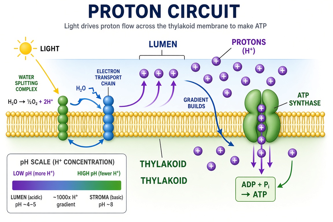
Verifique: lados lúmen vs estroma — os prótons acumulam-se dentro do lúmen; o ciclo de Calvin acontece no estroma, não no lúmen.
8. Resposta da planta à seca
A plant-physiology figure of drought response: roots sensing dry soil, abscisic
acid traveling up the xylem, guard cells closing stomata, leaf rolling and
osmoprotectant accumulation, with a healthy versus stressed plant pair.
Label "ABA", "GUARD CELL", "STOMATA", "OSMOPROTECTANT". Title: "SURVIVING DRYNESS".
Verifique: os estômatos fecham (células-guarda flácidas), não abrem, na planta estressada; a seta de ABA corre raiz → folha pelo xilema.
Genética e biologia molecular
9. Mecanismo CRISPR-Cas9
A gene-editing mechanism figure: the Cas9 protein as a bilobed clamp, guide RNA
threaded inside base-pairing with the target DNA, the HNH and RuvC domains cutting
both strands, and a repaired insert outcome panel. Label "CAS9", "GUIDE RNA",
"TARGET DNA", "CUT SITE", "INSERT". Title: "GENE EDITING".
Verifique: o RNA-guia pareia com a fita-alvo; a sequência PAM fica na fita não alvo, imediatamente após o corte.
10. Ligação anticorpo-antígeno
A molecular figure of an antibody with its two heavy and two light chains, the
antigen-binding sites gripping a small antigen, and one magnified inset of the
paratope-epitope interface. Label "HEAVY CHAIN", "LIGHT CHAIN", "PARATOPE",
"EPITOPE". Title: "ANTIBODY STRUCTURE".

11. Bacteriófago T4
A structural figure of the T4 bacteriophage with its icosahedral head, contractile
sheath, tail fibers and base plate, plus one inset showing the tail injecting DNA
into a bacterial envelope. Label "HEAD", "SHEATH", "TAIL FIBER", "BASE PLATE",
"DNA". Title: "T4 PHAGE".

Verifique: as fibras caudais aderem antes da contração da bainha, no detalhe da infecção.
12. Estágios de agregação do beta-amiloide
A four-stage molecular figure of peptide aggregation: free monomers, small oligomer
rings, a growing fibril strand shown as a twisted beta-sheet ladder, and a dense
senile plaque mass with microglia reacting at the edge. Label "MONOMER", "OLIGOMER",
"FIBRIL", "PLAQUE". Title: "AGGREGATION".
Verifique: a secção transversal da fibrila é uma pilha de folhas beta; se os degraus parecerem uma alfa-hélice, gere de novo.
Microbiologia e laboratório
13. Layout de placa de 96 poços
A top-down layout of a 96-well microplate set up for a dose-response assay: eight
rows by twelve columns, control wells in the outer columns, a serial dilution
gradient across the middle, and triplicate rows. Label "CONTROL", "DILUTION
SERIES", "REPLICATES" and add a small color-concentration legend.
Title: "PLATE LAYOUT".
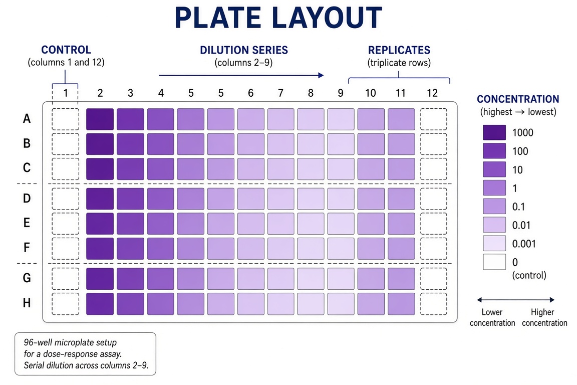
Verifique: o gradiente de diluição deve correr em um só sentido pelas colunas; as triplicatas são linhas, não colunas.
14. Fluxo de trabalho RT-qPCR
A bench workflow figure for a quantitative PCR run: template and master mix on ice,
plate setup with standards and controls in a layout grid, thermal cycling profile
with annealing step, and melt-curve verification at the end. Label "MASTER MIX",
"STANDARDS", "THERMAL CYCLE", "MELT CURVE". Title: "THE QPCR RUN".

15. ELISA sanduíche
A four-step laboratory workflow for a sandwich ELISA from coating the plate through
blocking and sample incubation to detection, each step as a small labeled panel
connected by arrows. Label "COATING", "BLOCKING", "INCUBATION", "DETECTION".
Title: "SANDWICH ELISA WORKFLOW".

Verifique: anticorpo de captura embaixo primeiro, anticorpo de detecção em cima — a ordem do sanduíche é o erro de laboratório didático mais comum.
Desenvolvimento e sistemas
16. Bioimpressão de um arcabouço tecidual
A figure of extrusion bioprinting: a printhead depositing parallel bioink strands
onto a grid scaffold, cells visible inside the strands in a magnified inset, and
the finished layered tissue construct on the right. Label "PRINTHEAD", "BIOINK",
"CELLS", "SCAFFOLD", "CONSTRUCT". Title: "BIOPRINTING".
17. Árvore genealógica das células do sangue
A classic hematopoiesis tree: the hematopoietic stem cell at the root branching
through progenitors into red cells, platelets, granulocytes, monocytes and
lymphocytes, each mature cell drawn at the leaf. Label "STEM CELL", "PROGENITOR",
"RED CELL", "PLATELET", "LYMPHOCYTE". Title: "BLOOD LINEAGES".
Oito variações rápidas
Troque o assunto, mantenha a gramática:
| # | Tema | Alteração em uma linha |
|---|---|---|
| 18 | Procarionte vs eucarionte | "A paired comparison of a bacterial cell and a eukaryotic cell… Label 'NUCLEOID', 'RIBOSOMES', 'PILI', 'NUCLEUS'" |
| 19 | Visão geral da mitose | "A five-panel row of mitosis stages: prophase, prometaphase, metaphase, anaphase, telophase… Label each panel" |
| 20 | Permuta na meiose | "A tetrad with chiasmata crossing over between homologous chromosomes… Label 'TETRAD', 'CHIASMA', 'RECOMBINANT'" |
| 21 | Cinética enzimática | "A lock-and-key figure plus a Michaelis-Menten curve inset… Label 'SUBSTRATE', 'ACTIVE SITE', 'VMAX', 'KM'" |
| 22 | Ciclo do nitrogênio | "A four-reservoir cycle figure: atmosphere, soil, plants, decomposers… Label 'N2', 'NITRIFICATION', 'ASSIMILATION', 'DENITRIFICATION'" |
| 23 | Sinapse neuronal | "A chemical synapse with vesicles fusing, neurotransmitters crossing the cleft, receptors on the postsynaptic side… Label 'VESICLE', 'CLEFT', 'RECEPTOR'" |
| 24 | Membrana celular | "A fluid-mosaic membrane cross-section with phospholipids, channel protein, glycoprotein… Label 'PHOSPHOLIPID', 'CHANNEL', 'GLYCOPROTEIN'" |
| 25 | Transcrição | "A DNA strand opening with RNA polymerase transcribing mRNA, one gene with promoter and terminator… Label 'PROMOTER', 'RNA POLYMERASE', 'mRNA'" |
Lista de verificação científica
- [ ] Todos os rótulos estão escritos corretamente?
- [ ] Todas as estruturas pedidas estão presentes?
- [ ] Posições e proporções são razoáveis frente à sua referência?
- [ ] As setas mostram a direção correta?
- [ ] As etapas aparecem na ordem correta?
- [ ] A figura foi conferida com uma fonte confiável?
Próximos passos
Execute qualquer prompt acima nas predefinições de biologia e verifique com seu livro didático antes da aula. Leituras mais profundas: prompts de anatomia para coração, cérebro e neurônios, prompts de ilustração botânica e os prompts de ilustração zoológica. Para o que costuma dar errado e como corrigir via prompt, veja 12 erros comuns de IA em ilustração científica.