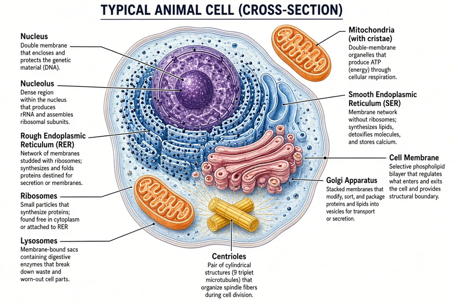
The best animal-cell diagram is matched to its audience. A middle-school overview may need the nucleus, cell membrane, cytoplasm, mitochondria, ribosomes, endoplasmic reticulum, Golgi apparatus, and lysosomes. A more advanced figure can distinguish rough and smooth ER, show nuclear pores, or explain vesicle traffic. Avoid packing every possible structure into one image. Ask for a separate unlabeled study version only after the reviewed labeled figure is correct, and remember that a typical teaching diagram is a simplified composite rather than a literal portrait of one cell.
Animal Cell Diagram — AI Generator
Create a labeled animal cell cross-section for a lesson, worksheet, presentation, or study guide. Specify the required organelles and audience, keep labels readable, and verify each structure instead of treating the generated image as an atlas plate.
- Match organelle count to the learner level
- Use leader lines that end on the intended structure
- Distinguish animal-cell features from plant-cell features
- Create an unlabeled study copy only after review
Prompt
Create a textbook-style cross-section of a typical animal cell. Label the cell membrane, cytoplasm, nucleus, nucleolus, nuclear envelope, mitochondria, rough endoplasmic reticulum, smooth endoplasmic reticulum, ribosomes, Golgi apparatus, lysosomes, vesicles, and centrosome. Use distinct but restrained colors, clear leader lines, short labels, generous spacing, and a white background. Do not add a cell wall, chloroplasts, or a large central vacuole. Treat the result as a simplified teaching model.
Scientific illustration resources
Prompt the subject, then verify the science
Use the matching prompt guide to refine this draft. Before publishing, check every label, structure and relationship against an authoritative source or a qualified subject expert.
Related images
More biology entries from the same generator
 Plant Cell Diagram — AI GeneratorView · biology
 Cell Diagram — AI GeneratorView · biology
 Photosynthesis Diagram — AI GeneratorView · biology
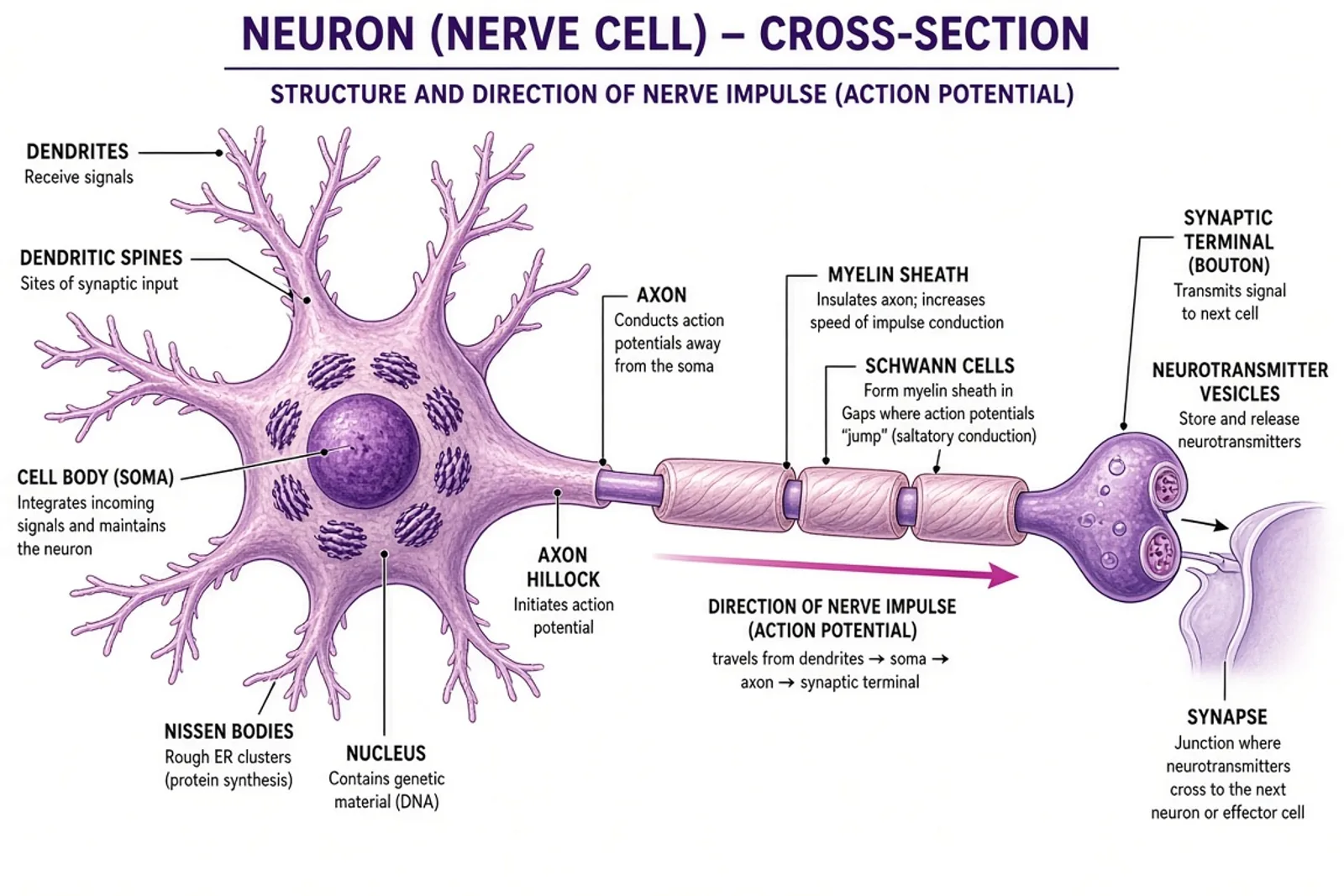 Neuron Diagram — AI GeneratorView · biology
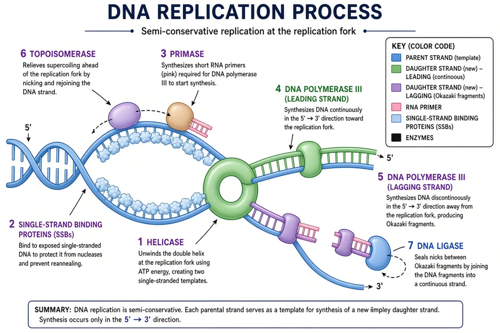 DNA Replication Diagram — AI GeneratorView · biology
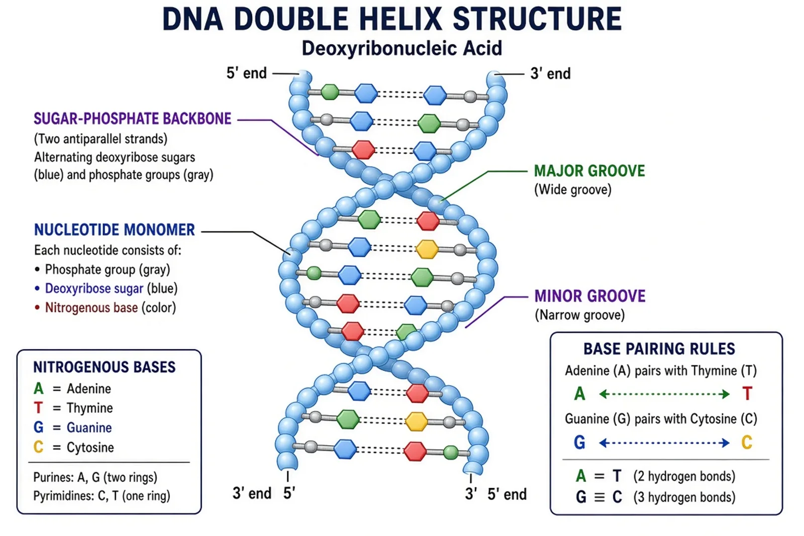 DNA Diagram — AI GeneratorView · biology
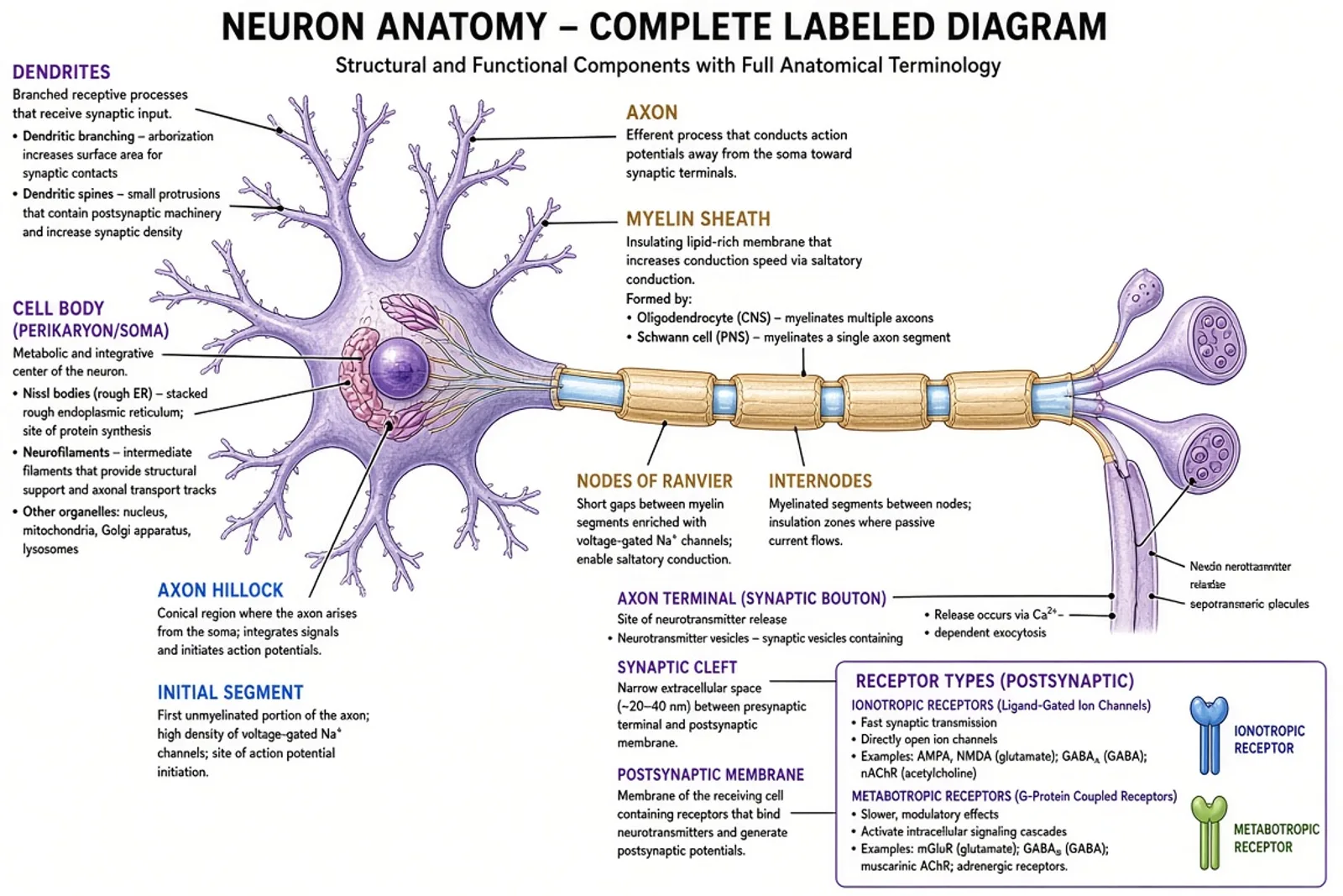 Labeled Neuron Diagram — AI GeneratorView · biology
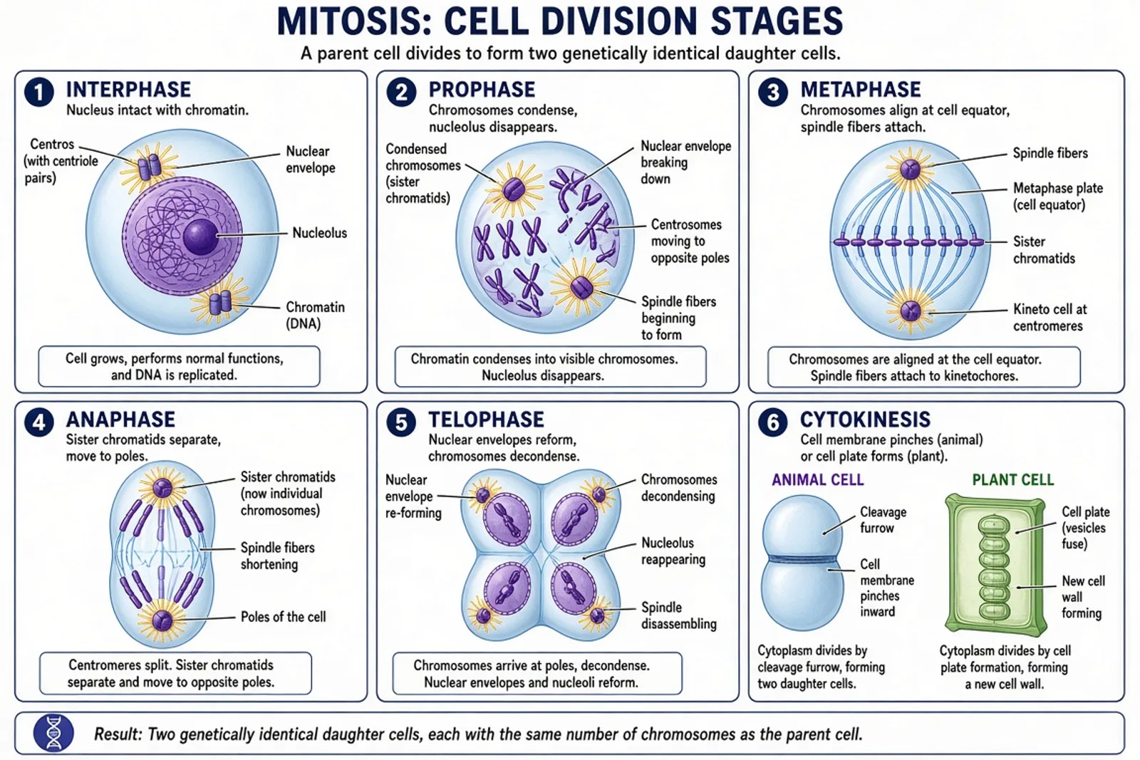 Cell Division Diagram — AI GeneratorView · biology
Frequently asked questions
Which organelles should a labeled animal cell include?
A practical overview includes the nucleus, cell membrane, cytoplasm, mitochondria, ribosomes, rough and smooth endoplasmic reticulum, Golgi apparatus, lysosomes, vesicles, and centrosome. Adjust the list to the course level.
How is an animal cell diagram different from a plant cell diagram?
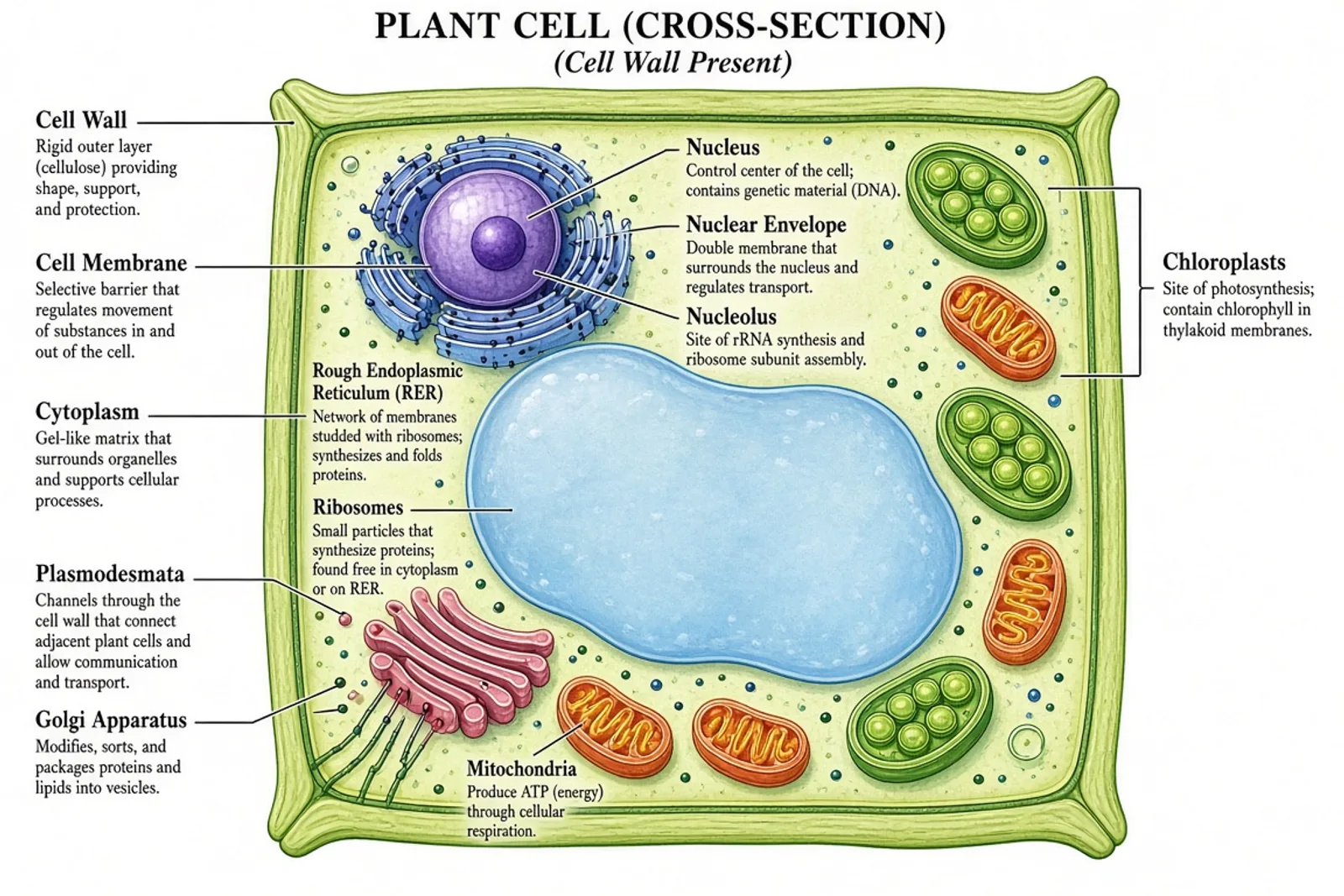
A typical animal-cell teaching diagram lacks a cell wall, chloroplasts, and one large central vacuole. Plant and animal cells share many organelles, so label the distinguishing structures carefully rather than relying only on shape.
Can I make a blank diagram for a labeling activity?
Yes. First verify the labeled version, then generate or prepare a matching copy without the terms. Keep leader lines or a word bank if they support the exercise, and retain the reviewed key for assessment.