This double helix — antiparallel strands, color-coded base pairs, groove callouts, a replication inset — came from a single 40-word prompt. Below are 25 biology prompts at that same standard, organized by topic, each with its label list and the specific thing to verify. Paste them straight into the generator or bend them to your exact topic.
Open the biology presets · Full workflow guide · 30 examples gallery
How to read this pack: every prompt follows one grammar — subject, then panels, then quoted labels, then a title. The quoted labels are what make text render correctly; the named panels are what make the layout come out as a figure instead of wall art. If a topic below doesn't match your lesson exactly, change the subject line and keep the structure.
Cells
1. DNA double helix
An elegant structural figure of the DNA double helix: two antiparallel strands,
base pairs as rungs with the four bases color-coded, a major and minor groove
callout, and one unzipping replication inset. Label "STRAND", "BASE PAIR", "MAJOR
GROOVE", "MINOR GROOVE". Title: "THE DOUBLE HELIX".
Verify: strands must run antiparallel (5'→3' one way, 3'→5' the other); A-T pairs have two hydrogen-bond bars, G-C three — check the inset didn't even them out.
2. DNA replication fork
A DNA replication fork with the parental double helix opening into a leading strand
and a lagging strand with Okazaki fragments, helicase at the fork and DNA polymerase
on the new strand, synthesis in the 5' to 3' direction. Label "HELICASE", "LEADING
STRAND", "LAGGING STRAND", "OKAZAKI FRAGMENT", "DNA POLYMERASE".
Title: "THE REPLICATION FORK".
Verify: the lagging strand's Okazaki fragments must point away from the fork; both new strands synthesize 5'→3' — if the drawing shows otherwise, regenerate with that constraint named.
3. Animal cell organelles
A large cutaway animal cell with its organelles arranged realistically, each
organelle shown once with a short functional caption beside it in a small grid
around the cell. Label "NUCLEUS", "MITOCHONDRION", "ROUGH ER", "GOLGI",
"LYSOSOME". Title: "THE ANIMAL CELL".
Verify: rough ER hugging the nucleus; centrosome near the nucleus, not at the membrane. Two cells compared:
4. Animal cell vs plant cell
A paired cutaway comparison of an animal cell and a plant cell at matching scale,
with shared organelles in the middle band and unique structures at the outer edges,
plus a small legend for the color coding. Label "NUCLEUS", "MITOCHONDRION", "CELL
WALL", "CHLOROPLAST", "VACUOLE". Title: "ANIMAL VS PLANT".

Verify: the large central vacuole and cell wall belong to the plant side only; check the color legend matches what's actually drawn.
5. Four specialized animal cells
A comparative row of four specialized cells at matching scale: a neuron with
dendrites and long axon, a red blood cell as a biconcave disc, a sperm cell, and a
macrophage spreading pseudopods, each with two functional labels.
Title: "CELL SPECIALISTS".
Verify: scale claims are dangerous here — flag that the row is not to scale (a red cell is ~7 µm, a neuron's axon can reach a meter) unless you add a scale bar.
Photosynthesis and plant biology
6. Photosynthesis light reactions (Z-scheme)
A Z-scheme figure of the light reactions: photosystem two splitting water and
sending electrons through plastoquinone, cytochrome, plastocyanin to photosystem
one, ferredoxin reducing NADP, and the proton gradient feeding ATP synthase.
Label "PSII", "PSI", "PLASTOQUINONE", "NADP", "ATP SYNTHASE".
Title: "Z SCHEME".
Verify: electron flow direction PSII → PSI; water is split at PSII (oxygen released there), ATP synthase sits in the thylakoid membrane with protons above it in the lumen.
7. Chloroplast proton traffic
A chloroplast figure centered on the thylakoid membrane: protons pumped into the
lumen by the electron transport chain flowing back out through ATP synthase,
stroma-side NADP reduction, and one granum stack inset showing where all this sits.
Label "LUMEN", "STROMA", "PROTON", "ATP SYNTHASE", "NADP".
Title: "PROTON TRAFFIC".
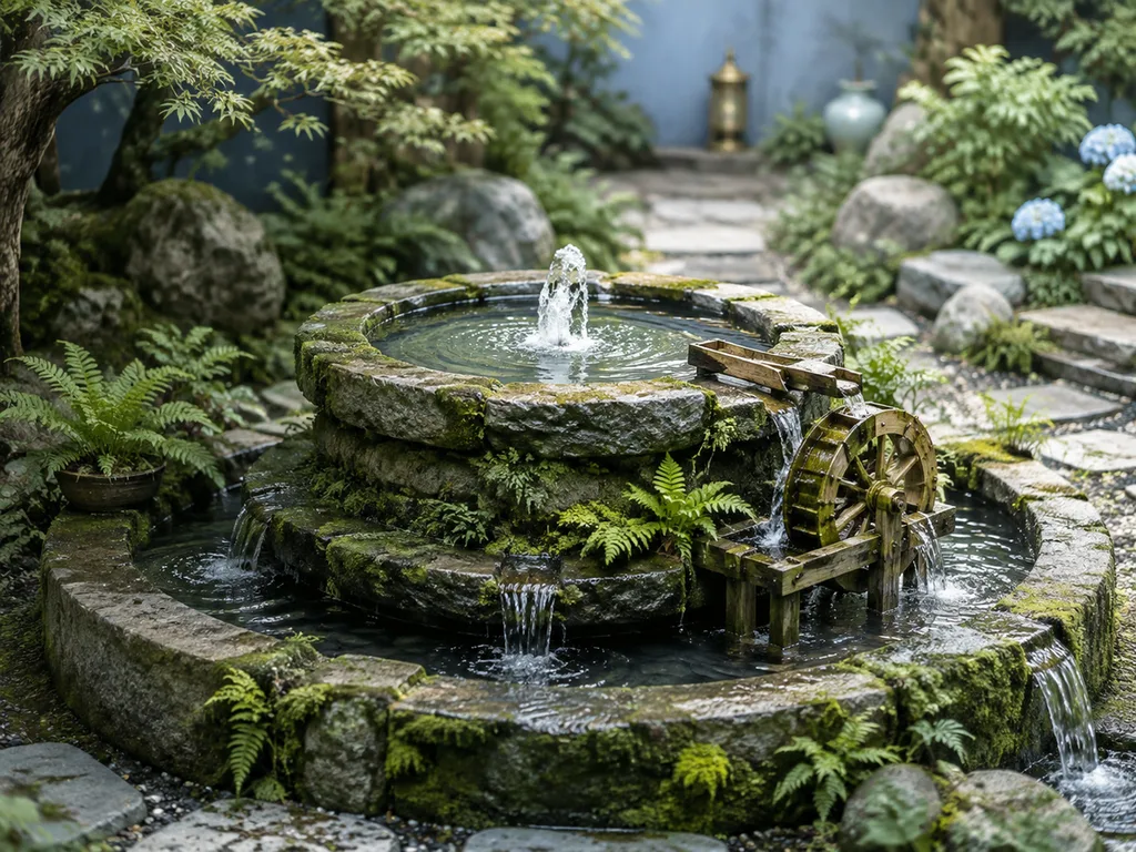
Verify: lumen vs stroma sides — protons accumulate inside the lumen; the Calvin cycle belongs in the stroma, not the lumen.
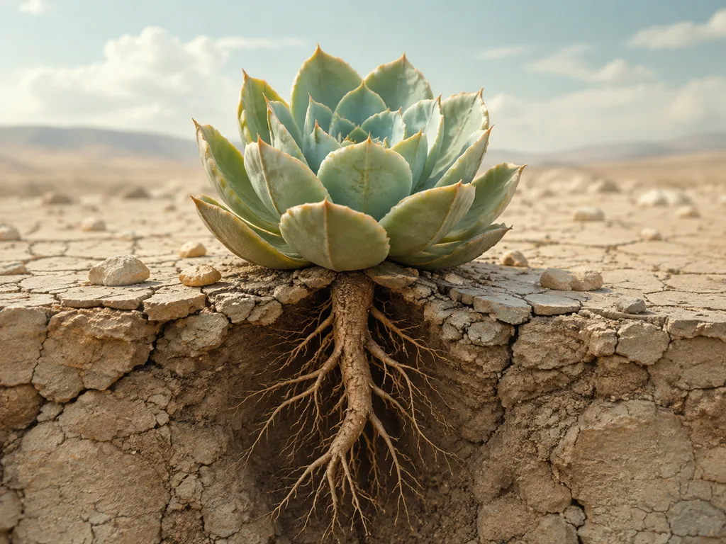
8. Plant drought response
A plant-physiology figure of drought response: roots sensing dry soil, abscisic
acid traveling up the xylem, guard cells closing stomata, leaf rolling and
osmoprotectant accumulation, with a healthy versus stressed plant pair.
Label "ABA", "GUARD CELL", "STOMATA", "OSMOPROTECTANT". Title: "SURVIVING DRYNESS".
Verify: stomata close (guard cells flaccid), not open, in the stressed plant; ABA arrow runs root → leaf through xylem.
Genetics and molecular biology
9. CRISPR-Cas9 mechanism
A gene-editing mechanism figure: the Cas9 protein as a bilobed clamp, guide RNA
threaded inside base-pairing with the target DNA, the HNH and RuvC domains cutting
both strands, and a repaired insert outcome panel. Label "CAS9", "GUIDE RNA",
"TARGET DNA", "CUT SITE", "INSERT". Title: "GENE EDITING".
Verify: the guide RNA base-pairs with the target strand; the PAM sequence sits on the non-target strand immediately after the cut.
10. Antibody-antigen binding
A molecular figure of an antibody with its two heavy and two light chains, the
antigen-binding sites gripping a small antigen, and one magnified inset of the
paratope-epitope interface. Label "HEAVY CHAIN", "LIGHT CHAIN", "PARATOPE",
"EPITOPE". Title: "ANTIBODY STRUCTURE".
11. Bacteriophage T4
A structural figure of the T4 bacteriophage with its icosahedral head, contractile
sheath, tail fibers and base plate, plus one inset showing the tail injecting DNA
into a bacterial envelope. Label "HEAD", "SHEATH", "TAIL FIBER", "BASE PLATE",
"DNA". Title: "T4 PHAGE".
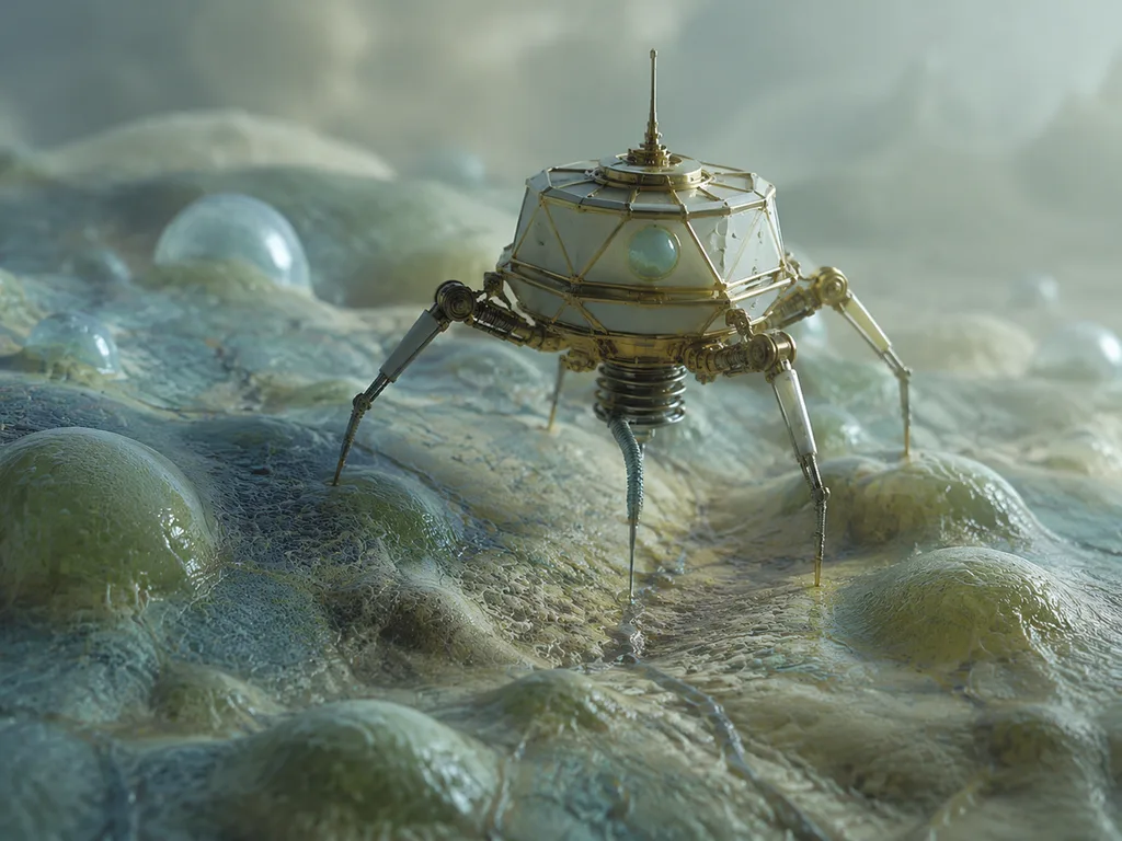
Verify: tail fibers attach before sheath contraction in the infection inset.
12. Amyloid-beta aggregation stages
A four-stage molecular figure of peptide aggregation: free monomers, small oligomer
rings, a growing fibril strand shown as a twisted beta-sheet ladder, and a dense
senile plaque mass with microglia reacting at the edge. Label "MONOMER", "OLIGOMER",
"FIBRIL", "PLAQUE". Title: "AGGREGATION".
Verify: the fibril cross-section is a beta-sheet stack; if the rungs look like an alpha helix, regenerate.
Microbiology and lab
13. 96-well plate layout
A top-down layout of a 96-well microplate set up for a dose-response assay: eight
rows by twelve columns, control wells in the outer columns, a serial dilution
gradient across the middle, and triplicate rows. Label "CONTROL", "DILUTION
SERIES", "REPLICATES" and add a small color-concentration legend.
Title: "PLATE LAYOUT".
Verify: dilution gradient must run one direction across columns; triplicates are rows, not columns.
14. RT-qPCR workflow
A bench workflow figure for a quantitative PCR run: template and master mix on ice,
plate setup with standards and controls in a layout grid, thermal cycling profile
with annealing step, and melt-curve verification at the end. Label "MASTER MIX",
"STANDARDS", "THERMAL CYCLE", "MELT CURVE". Title: "THE QPCR RUN".

15. Sandwich ELISA
A four-step laboratory workflow for a sandwich ELISA from coating the plate through
blocking and sample incubation to detection, each step as a small labeled panel
connected by arrows. Label "COATING", "BLOCKING", "INCUBATION", "DETECTION".
Title: "SANDWICH ELISA WORKFLOW".
Verify: capture antibody down first, detection antibody on top — the sandwich order is the most common classroom error.
Development and systems
16. Bioprinting a tissue scaffold
A figure of extrusion bioprinting: a printhead depositing parallel bioink strands
onto a grid scaffold, cells visible inside the strands in a magnified inset, and
the finished layered tissue construct on the right. Label "PRINTHEAD", "BIOINK",
"CELLS", "SCAFFOLD", "CONSTRUCT". Title: "BIOPRINTING".
17. Blood cell family tree
A classic hematopoiesis tree: the hematopoietic stem cell at the root branching
through progenitors into red cells, platelets, granulocytes, monocytes and
lymphocytes, each mature cell drawn at the leaf. Label "STEM CELL", "PROGENITOR",
"RED CELL", "PLATELET", "LYMPHOCYTE". Title: "BLOOD LINEAGES".
Eight quick variants
Swap the subject, keep the grammar:
| # | Topic | One-line change |
|---|---|---|
| 18 | Prokaryote vs eukaryote | "A paired comparison of a bacterial cell and a eukaryotic cell… Label 'NUCLEOID', 'RIBOSOMES', 'PILI', 'NUCLEUS'" |
| 19 | Mitosis overview | "A five-panel row of mitosis stages: prophase, prometaphase, metaphase, anaphase, telophase… Label each panel" |
| 20 | Meiosis crossover | "A tetrad with chiasmata crossing over between homologous chromosomes… Label 'TETRAD', 'CHIASMA', 'RECOMBINANT'" |
| 21 | Enzyme kinetics | "A lock-and-key figure plus a Michaelis-Menten curve inset… Label 'SUBSTRATE', 'ACTIVE SITE', 'VMAX', 'KM'" |
| 22 | Nitrogen cycle | "A four-reservoir cycle figure: atmosphere, soil, plants, decomposers… Label 'N2', 'NITRIFICATION', 'ASSIMILATION', 'DENITRIFICATION'" |
| 23 | Neuron synapse | "A chemical synapse with vesicles fusing, neurotransmitters crossing the cleft, receptors on the postsynaptic side… Label 'VESICLE', 'CLEFT', 'RECEPTOR'" |
| 24 | Cell membrane | "A fluid-mosaic membrane cross-section with phospholipids, channel protein, glycoprotein… Label 'PHOSPHOLIPID', 'CHANNEL', 'GLYCOPROTEIN'" |
| 25 | Transcription | "A DNA strand opening with RNA polymerase transcribing mRNA, one gene with promoter and terminator… Label 'PROMOTER', 'RNA POLYMERASE', 'mRNA'" |
Scientific review checklist
- [ ] Are all labels spelled correctly?
- [ ] Are all requested structures present?
- [ ] Are positions and proportions reasonable against your reference?
- [ ] Do arrows show the correct direction?
- [ ] Are stages shown in the correct order?
- [ ] Has it been checked against a trusted source?
Next steps
Run any prompt above in the biology presets, then verify against your textbook before class. Deeper reads: anatomy prompts for heart, brain and neurons, botanical prompts, and the animal illustration guide. For what generally goes wrong and how to fix it by prompt, see 12 common AI scientific illustration errors.
<!-- scientific-illustration-cluster-links:start -->
Continue through the scientific illustration collection
Create a scientific illustration · Browse the scientific illustration resource hub
Related: 20 Human Anatomy Diagram Prompts for Heart, Brain and Neurons · 20 Botanical Illustration Prompts for Flowers, Leaves and Life Cycles
<!-- scientific-illustration-cluster-links:end -->