LIFE SCIENCES
Medical Anatomy & Physiology
Organ cutaways, tissue layers, and blood-flow direction with standard anatomical labels.
- Organ cutaways
- Blood flow
Text to Clinical Figure · Medical Recipe
Generate clinical-style medical figures with color-coded anatomy, flow direction, and standard terminology — organ cutaways, surgical approaches, and mechanism diagrams for education and communication.
Use this workflow for a medical visual deliverable: a labeled organ, mechanism or patient-education draft. It is separate from career content about medical illustrators and always keeps qualified review in the production loop.

LIFE SCIENCES
Organ cutaways, tissue layers, and blood-flow direction with standard anatomical labels.
LIFE SCIENCES
Molecular pathways and cell machinery for mechanism figures.
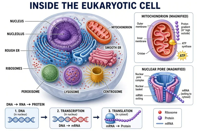
Made with GLMImage
Inspect real clinical-style drafts and use their structure as a starting point—not as unreviewed medical truth.
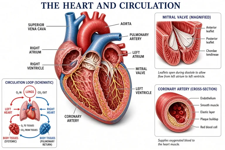
 Human heart blood flow (validation)
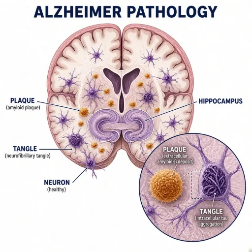 Alzheimer pathology in the hippocampus
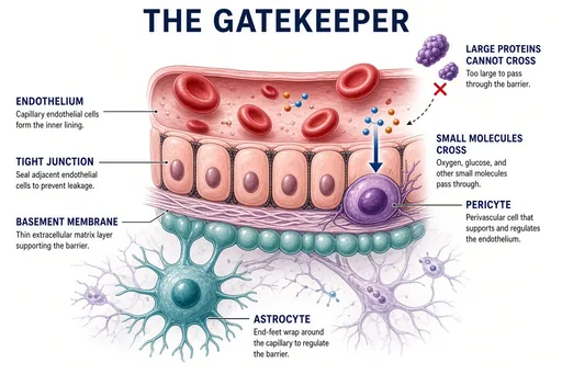 Blood-brain barrier concept
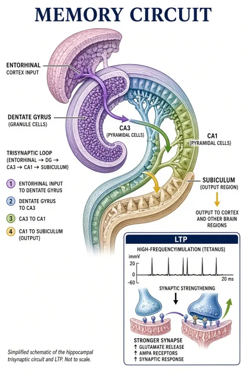 Hippocampus memory circuit
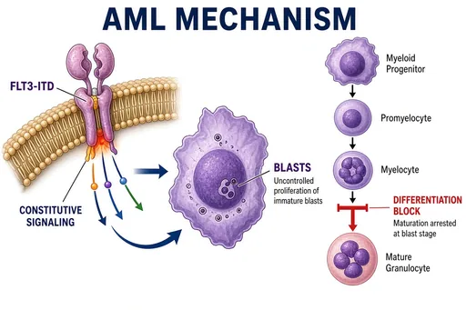 AML FLT3-ITD pathogenesis
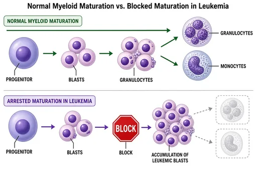 Myeloid differentiation block
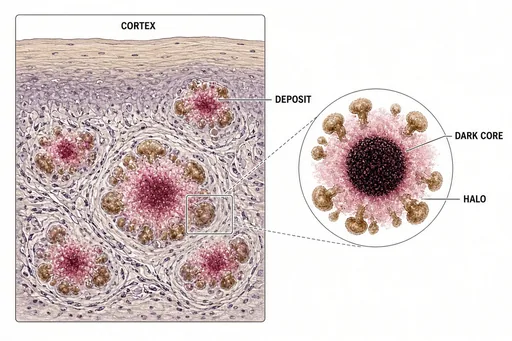 Plaque burden in brain tissue
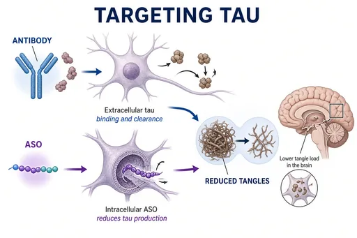 Anti-tau therapeutic strategies
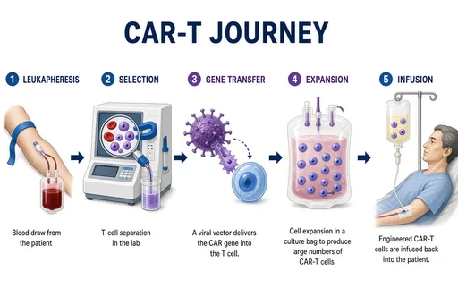 CAR-T manufacturing workflow
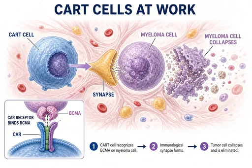 BCMA CAR-T in myeloma
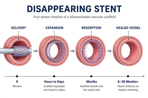 Biodegradable stent lifecycle
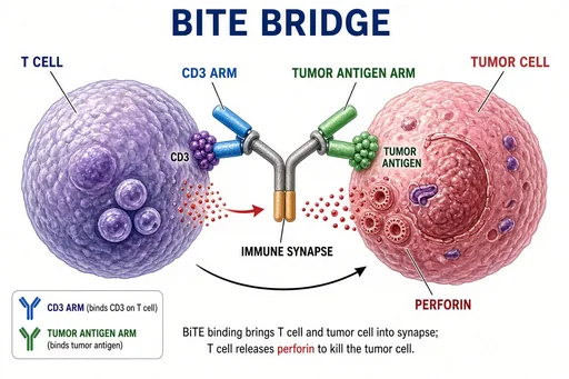 Bispecific antibody bridging
Focused workflow
Organ, region, or mechanism — plus the orientation you need.
Exact terms in quotation marks; ask for arrows where direction matters.
Check against a standard atlas; correct one element per iteration.
A qualified reviewer signs off before patient-facing or published use.
Practical starting points
Use these as structures, not magic phrases. Replace the subject, purpose and constraints with details from your own project.
Create a labeled diagram of a nephron showing the glomerulus, Bowman capsule, proximal tubule, loop of Henle, distal tubule, and collecting duct, with filtrate flow arrows, clinical textbook style.
Mechanism · physiology
Create a sagittal section of the knee joint labeling the "femur", "tibia", "patella", "meniscus", "anterior cruciate ligament", and "posterior cruciate ligament".
Regional anatomy · sagittal
Create a four-panel medical illustration of wound healing stages — hemostasis, inflammation, proliferation, remodeling — each panel labeled with the key cell types.
Process panels · patient education
Medical illustration covers everything from textbook anatomy to the mechanism diagram in a drug ad. The medical recipe renders clean clinical-style figures: neutral backgrounds, color-coded structures, leader-line labels using standard terminology, and directional cues for flow.
Typical uses are teaching decks, patient-education handouts, thesis figures, and health-content marketing — anywhere a correct, readable anatomical figure is needed faster than manual illustration.
Every generated medical figure is a draft. Verify anatomy against a standard atlas, confirm labels and laterality, and have a qualified clinician or medical illustrator review anything shown to patients or published. The generator accelerates layout and labeling; it does not replace clinical review.
Avoid diagnostic or treatment claims in the surrounding copy, and never present an AI figure as a scan or real patient image.
Guides for medical and anatomical illustration.
Yes — describe the approach and what should be visible. Treat surgical figures as educational drafts; verify orientations and relationships before use in teaching materials.
No. It removes the layout and labeling grind so illustrators and educators start from a strong draft; final published figures still need professional review.
Standard clinical and anatomical terminology — chambers, valves, tubule segments, ligament names. Put exact wording in quotation marks so terms render correctly.
Yes, as drafts that a qualified clinician reviews first. Verify anatomy, labels, and laterality against a standard atlas, and never present generated figures as scans or real patient images.
Specify the anatomical view, required labels and flow direction. Use the result as a draft, then verify it against an authoritative atlas.
Draft a medical illustration